Magnetic Resonance Imaging (MRI)
The MRI is an examination in radiologic and nuclear medical diagnostics. In medical imaging, magnetic fields and radio waves are created to produce tomographic images of the human body. This allows an assessment of the structure and function of tissues and organs in the body and therefore also many pathologic changes. This process does not use radiation.
Other names: Magnetic resonance tomography (MRT 1.5 Tesla)
What is a Magnetic Resonance Imaging (MRI)?
Magnetic resonance imaging (MRI) is an examination method in radiology and nuclear medicine creating images of internal organs. In contrast to an X-ray or computed tomography, X-rays magnetic fields and radio waves are used rather than x-rays.
How does magnetic resonance imaging (MRI) work?
Basis of the MRI are very strong magnetic fields, in which patients are examined. By electromagnetic fields in the form of radio waves in the VHF frequency range, certain atomic nuclei (hydrogen nuclei) are stimulated selectively in the body utilizing the fact that there are many hydrogen atoms are present in the human body. The MRI device records the physical response of the excited nuclei. A powerful computer converts signals by a very complex calculation method and combines them to very detailed 2 - or 3-dimensional images. These can then be evaluated by a physician. The images show particularly well the condition of the soft tissues or organs of the body and joints and provide information on their structure and function. Tissue having a high water content and fat are light colored while tissue with low water content is darker.
For which indication is the method useful?
Practically all areas of the body can be imaged by the MRI:
- Brain and cranium
- Facial and Eye
- Spine and spinal cord
- Soft tissues of the chest (for the lungs, currently the computed tomography is still more appropriate.)
- Upper abdomen and pelvic organs
- All joints, particularly with respect to cartilage, ligaments and capsule structures
- Muscle processes
- Vessels by means of a special technique, the so-called MR angiography
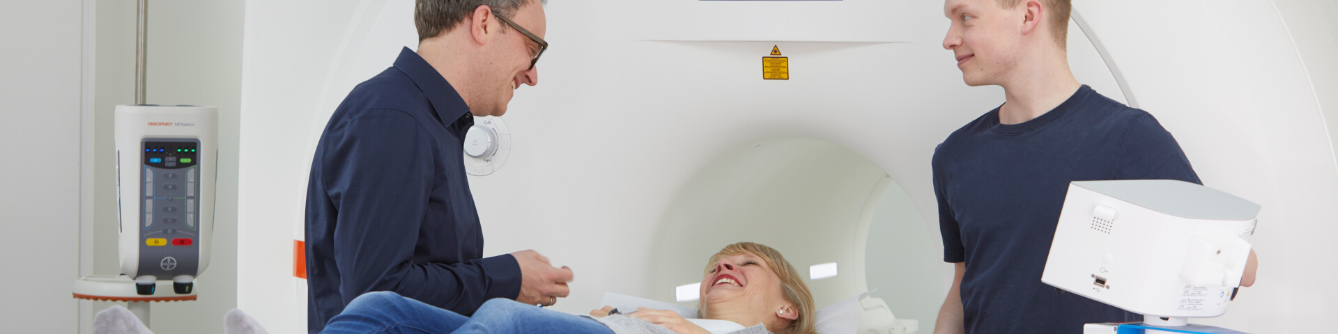
Equipment technology
So-called "closed devices" still have a greater advantage over open systems because they allow stronger magnetic fields. This is an important factor for the image quality and scan speed. Our unit of the brand Siemens Symphony Quantum (field strength of 1.5 Tesla) has the form of a short Tunnel of 60cm in diameter and open at the front and the back, meaning it is well lit and ventilated. At the systematic variation of the magnetic field (see "What is it?") very loud sometimes compressed air-hammer-like noise are caused by a so-called gradient. Unfortunately currently the rule of thumb: "The louder the noise, the better the device" is still dominating. This noise level is counteracted with suitable hearing protection. However, the patient should be carefully monitored during the exam. The patient can also use an "emergency bell" any time during the exam. Depending on the medical issue and the body region to be examined, we use different "antennas"/coils.