
The Spine – basic functions, anatomy and disorders
As long as we can move around without pain and participate in sports without problems, we are hardly aware of the complex workings of our body. The smooth interaction of the joints, but particularly the painless and unrestricted functioning of our central axial skeleton, we take the mobility of our spine for granted. However, disorders and diseases in this system are likely to substantially affect t the quality of life greatly.
The spine has two extreme important functions
Firstly, it allows movements of the fuselage in all directions and effectively absorbs the weight resulting from our movements. Although, it is flexible, it is very strong. In everyday life – but especially during sports and physical activities – the spine is already exposed to an enormous amount of stress. Nevertheless, because of its unique construction it can withstand this stress effectively. For example, the cartilage layer of the vertebral joints is compared to the size of the joint, the thickest articular cartilage in the body. This means that the spine can also withstand substantial forces and weight.
The second major function of the spine is the protection it offers to the spinal cord running in the spinal canal and the exiting nerve roots. Just as the skull does for the brain, the spinal column forms a bony armor around the spinal cord.
Since the spine must be stable and flexible at the same time, a particularly ingenious design is necessary. Only a perfectly coordinated interplay of vertebrae, intervertebral discs, muscles and ligaments enables us to move head and trunk in all directions, as well as to withstand high loads in everyday life and especially in sports or during physical work. We only feel how sensitive our spine is when diseases or disorders occur in this system and massively impair our quality of life.
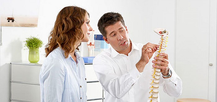
How is the spine constructed?
The spine consists of 24 bony building blocks, the vertebral bodies, which are strung together like a chain. They transfer the weight of the upper body to the pelvis and give the spine its stability. 7 vertebrae form the cervical, 12 the thoracic and 5 the lumbar spine. This is followed by the sacrum and coccyx (structure of the spine). While the spine forms a straight line from behind, when viewed from the side it has the shape of a double S. The cervical and lumbar spine are convex to the front (lordosis), the thoracic spine and the sacrum are arched backwards (kyphosis). Shocks and running movements are effectively cushioned, vibrations are attenuated and evenly distributed over the body.
Facet joints - Small joints with great function
In addition, each vertebral arch has two paired articular processes. The so-called facet joints (intervertebral joints) connect the vertebral arches of two adjacent vertebrae and control their mobility. Almost a quarter of the total load is absorbed by the facet joints, which is why their cartilage is particularly developed. But as with any other joint in our body, the cartilage of the facet joints wears over time and arthrosis can develop, the so-called facet joint arthrosis.
Intervertebral discs - The natural shock absorbers
The intervertebral discs lie between the individual vertebral bodies like a kind of buffer. They contribute to mobility, absorb daily stress and distribute the load evenly across the spine.
Each intervertebral disc consists of an inner gelatinous nucleus (nucleus pulposus) and an outer fibrous ring (anulus fibrosus) of collagenous connective tissue. Its coarse fibres are crossed so that excessive movements are prevented and optimum power transmission is made possible. The gelatinous core, on the other hand, consists largely of protein molecules that can store water excellently. Like a sponge, the disc absorbs fluid and nutrients from its surroundings and thus serves as a natural shock absorber between the bony vertebrae. If pressure is exerted on the intervertebral discs when the spine is under strain (e.g. standing or sitting for a long time), they lose fluid and become thinner. When relieved, e.g. at night or when walking evenly, the intervertebral discs absorb water and nutrients again. The supply of the intervertebral discs deteriorates with age. They lose fluid and elasticity and thus part of their buffering effect.
How is the spine structured
The spine is divided into five sections: Cervical, thoracic and lumbar spine, sacrum and coccyx.
Cervical spine
The cervical spine consists of 7 cervical vertebrae and is the most mobile part of the spine. As a result, your intervertebral discs are exposed to great strain and a herniated disc or wear and tear (cervical spine syndrome) can occur more frequently in this section. The uppermost cervical vertebra (atlas) differs in its construction plan from the remaining vertebrae. Instead of a vertebral body, it consists of an annular bone arch which, together with the 2nd cervical vertebra (axis), forms a kind of swivel joint. This special construction makes up and down movements as well as the rotation of our head possible.
Thoracic spine
The thoracic spine consists of 12 vertebrae to which the ribs are attached. Each rib is connected to the transverse process of a thoracic vertebra via a small joint, which enables the breathing movement of our thorax. In contrast to the cervical spine, the thoracic spine allows only little movement. Herniated discs are therefore rather rare in this part of the spine.
Lumbar spine
The thoracic spine is connected to 5 lumbar vertebrae. In the lumbar spine the bone substance, but also the strain is greatest, so that degenerative changes and complaints such as a herniated disc or a herniated disc usually occur there most frequently.
Sacrum
The next 5 vertebrae are fused into a single, wedge-shaped bone, the sacrum. The sacrum forms the pelvic belt together with the iliac bone, ischial bone and pubis.
Coccyx
The coccyx, which adjoins the lower sacrum, consists of 3 to 5 (usually 4) also fused vertebrae and is indispensable for the statics of the pelvis. This is where tendons and ligaments, especially of the pelvic floor and hips, come into play.